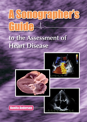
A Sonographer's Guide to the Assessment of Heart Disease
A Sonographer's Guide to the Assessment of Heart Disease
NON-RETURNABLE
Editor: Bonita Anderson
Publisher: Echotext
Published 2013-11
Hardcover, 507 pages
ISBN: 9780992322205
Reviews:
Doody's Review Service 4-Star Rating- Scored 94 out of 100.
About:
Written by a sonographer for sonographers, this text primarily discusses the role of echocardiography in the assessment of heart diseases. The book is mostly designed for students of echocardiography, teachers of echocardiography and cardiac sonographers working in routine clinical practice, but will also be very useful to echocardiologists and cardiac registrars. The goal of the text is to provide a comprehensive review of transthoracic echocardiography in the assessment of various cardiac pathologies. Refresher notes on cardiac anatomy and the relevant cardiac physiology and pathophysiology are included to expand the cardiac sonographer's knowledge in this area and further their understanding of various diseases, disease processes and associated findings. This comprehensive text is supported by over 720 figures including over 1,200 echo images, over 130 pathological photos and many illustrations. It also includes several appendices, numerous tables and technical tips which highlight key concepts. Hard-cover bound with 507 printed pages, this text is ready to be adopted as any sonographer's constant companion while dealing with the many challenges that arise in performing an echocardiographic examination.
Real-time images which correspond to figures in the book are now available on www.echotext.info.
About the Author:
Author of the popular Echocardiography: the Normal Examination and Echocardiographic Measurements, Bonita Anderson is currently Senior Lecturer in Cardiac Ultrasound at the Queensland University of Technology, Brisbane, Australia. Bonita has over 30 years experience as a sonographer and educator in echocardiography at The Prince Charles Hospital, also in Brisbane. An Accredited Medical Sonographer, Bonita holds a Diploma in Medical Ultrasonography (Cardiac) and a Master of Applied Science (Medical Ultrasound). A popular international lecturer in echocardiography, Bonita is often complimented on her ability to explain complex concepts in an easy-to-understand manner.
Table of Contents:
Chapter 1: Introduction to Basic Haemodynamic Calculations
Determination of Pressure Gradients
Volumetric Flow Calculations
Continuity Principle
Key Points
Further Reading
Chapter 2: Ventricular Size and Systolic Function
Ventricular Anatomy
Basic Cardiac Physiology
Measurements of the Left Ventricular Size
Measurements of Left Ventricular Systolic Function
Measurements of the Right Ventricular Size
Measurements of Right Ventricular Systolic Function
Key Points
Further Reading
Chapter 3: Ventricular Diastolic Function
Basic Principles of Diastole
Echocardiographic Parameters for Assessing Left Ventricular Diastolic Function
Echocardiographic Parameters for Assessing Left Ventricular Filling Pressures
Echocardiographic Parameters for Assessing Right Ventricular Diastolic Function
Echocardiographic Parameters for Identifying Increased Right Atrial Pressures
Grading of Right Ventricular Diastolic Function
Key Points
Further Reading
Chapter 4: Hypertensive Heart Disease
Systemic Hypertension
Pulmonary Hypertension
Key Points
Further Reading
Chapter 5: Ischaemic Heart Disease
Coronary Artery Anatomy
Coronary Artery Disease
Role of Echocardiography in Ischaemic Heart
Disease
Role of Stress Echocardiography in Ischaemic Heart Disease
Complications of Myocardial Infarction
Direct Imaging of Coronary Arteries
Non-Coronary Artery Diseases Causing Ischaemic Chest Pain
Non-Ischaemic Causes of Chest Pain
Key Points
Further Reading
Chapter 6: Cardiomyopathies
Definition and Classifications of Cardiomyopathies
Dilated Cardiomyopathy
Hypertrophic Cardiomyopathy
Restrictive Cardiomyopathy
Arrhythmogenic Right Ventricular Cardiomyopathy
Left Ventricular Non-Compaction
Key Points
Further Reading
Chapter 7: Aortic Valve Disease
Anatomy of the Aortic Valve and Aortic Root
Aortic Stenosis
Aortic Regurgitation
Key Points
Further Reading
Chapter 8: Mitral Valve Disease
Anatomy of the Mitral Valve
Mitral Stenosis
Mitral Regurgitation
Key Points
Further Reading
Chapter 9: Tricuspid and Pulmonary Valve Disease
Anatomy of the Tricuspid Valve
Echo Imaging of the Tricuspid Valve
Tricuspid Stenosis
Tricuspid Regurgitation
Echo Imaging of the Tricuspid Valve
Echo Imaging of the Pulmonary Valve
Pulmonary Stenosis
Pulmonary Regurgitation
Key Points
Further Reading
Chapter 10: Prosthetic Heart Valves
Basic Concepts
Role of Echocardiography
Haemodynamic Assessment of Prosthetic Heart Valves
Complications of Prosthetic Valves
Key Points
Further Reading
Chapter 11: Diseases of the Aorta
Anatomy and Structure of the Aorta
2D Imaging and Measurements of the Aorta
Aortic Aneurysms
Sinus of Valsalva Aneurysms
Acute Aortic Syndromes
Aortic Dissections
Traumatic Aortic Injury
Key Points
Further Reading
Chapter 12: Pericardial Disease
Anatomy and Function of the Pericardium
Pericardial Cysts
Congenitally Absent Pericardium
Acute Pericarditis
Pericardial Effusions
Cardiac Tamponade
Constrictive Pericarditis
Effusive-Constrictive Pericarditis
Key Points
Further Reading
Chapter 13: Infective Endocarditis and Cardiac Masses
Infective Endocarditis
Cardiac Tumours
Intracardiac Thrombus
Normal Anatomy Simulating Cardiac Masses
Imaging Artefacts Simulating Cardiac Masses
Key Points
Further Reading
Chapter 14: Systemic Diseases with Cardiac Manifestations
Introduction
Systemic Rheumatic Diseases
Hereditary Connective Disorders
Endocrine Disorders
Haematologic Disorders
Infiltrative Disorders
Storage Disorders
Hereditary Neuromuscular Disorders
Key Points
Further Reading
Chapter 15: Introduction to Congenital Heart Disease
Introduction
Shunt Lesions
Obstructive Lesions
Cyanotic Lesions
Key Points
Further Reading
Appendices
Appendix 1: Normal Haemodynamic Values for Selected Prosthetic Aortic Valves
Appendix 2: Normal Haemodynamic Values for Selected Prosthetic Mitral Valves
Appendix 3: Normal Haemodynamic Values for Selected Prosthetic Tricuspid Valves
Appendix 4: Normal Haemodynamic Values for Selected Prosthetic Pulmonary Valves
Appendix 5: Selected Congenital and Genetic Syndromes associated with Congenital Heart Disease
Appendix 6: Selected Surgeries for Various Congenital Heart Lesions
Appendix 7: Echocardiographic Parameter Tables
Glossary
Index

