
Videos from Interventional Cardiac Electrophysiology
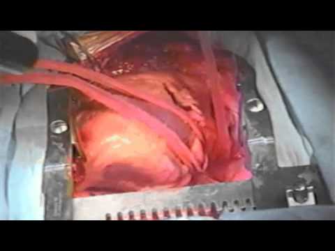
Video 66A.1

Video 66A.1 Typical right atrial argon cryothermia lesions as part of biatrial ablation procedure.
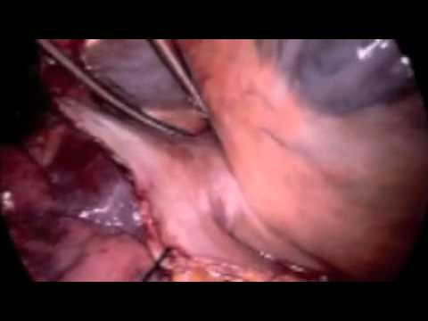
Video 66A.2
Video 66A.2 Sternotomy exposure of the left pulmonary veins off bypass.
Video 66A.3
Video 66A.3 Port access right pulmonary vein isolation using dry bipolar radiofrequency clamp.
Video 66A.4
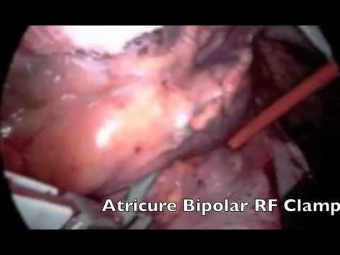
Video 66A.4 Port access left pulmonary vein isolation using dry bipolar radiofrequency clamp.
Video 66A.5

Video 66A.5 Port access left atrial appendectomy with pericardium reinforced stapler.
Video 66A.6

Video 66A.6 Synopsis of pericardioscopic procedure.
Video 67.1

Video 67.1 3D EnSite array balloon mapping of high right atrial pacing in a patient with refractory atrial flutter and atrial fibrillation before cavotricuspid isthmus ablation.
Video 67.2

Video 67.2 3D EnSite array balloon mapping of coronary sinus ostial pacing in a patient with refractory atrial flutter and AF before cavotricuspid isthmus ablation. Note that the left image shows a right lateral view and the right image shows a left anterior oblique view of the activation wavefront as it arises inferiorly at the posterior right atrium near the ostium.
Video 67.3

Video 67.3 3D EnSite array balloon mapping of dual-site right atrial pacing in a patient with refractory atrial flutter and AF before cavotricuspid isthmus ablation. Note that the left image shows a right lateral view and the right image shows a left anterior oblique view.
Video 67.4

Video 67.4 3D EnSite array balloon mapping of dual-site right atrial pacing in a patient with refractory atrial flutter and AF. Cavotricuspid isthmus linear ablation lesions and failure of paced wavefront to cross the linear lesion created by ablation. The wavefront leading edge is shown by the motion of the red asterisk, and it reflects off the line and propagates posteriorly.
Video 67.5A
Video 67.5A 3D EnSite array balloon mapping of propagation of atrial paced in a patient with refractory AF after right atrial compartmentalization. Linear lesions are created with radiofrequency ablation in the lateral right atrium (intercaval line), superior vena cava to fossa ovalis to coronary sinus ostium to tricuspid valve (septal line) and cavotricuspid isthmus line (isthmus) line. A: The 3D map is shown in the anterior projection and propagation of a paced wavefront in the anterior compartment is shown. Note that the wavefront remains confined to the anterior right atrial compartment and reflects off the linear lesions.
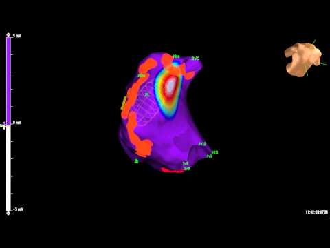
Video 67.5B

Video 67.5B 3D EnSite array balloon mapping of propagation of atrial paced in a patient with refractory AF after right atrial compartmentalization. Linear lesions are created with radiofrequency ablation in the lateral right atrium (intercaval line), superior vena cava to fossa ovalis to coronary sinus ostium to tricuspid valve (septal line) and cavotricuspid isthmus line (isthmus) line. B: The 3D map is shown in the posterocaudal projectionand propagation of a paced wavefront in the posterior
compartment is shown. Note that the wavefront arises at the coronary sinus ostial region and remains confined to the posteroseptal right atrial compartment and reflects off the linear lesions.